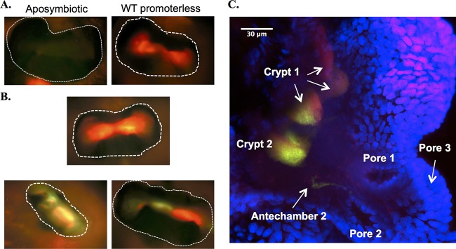
FIG 7.
Expression of PhoB-dependent reporters in symbiotic V. fischeri cells. (A and B) Juvenile squid were left uninoculated and aposymbiotic (A, left) or were infected with V. fischeri containing either the promoterless vector (pJLS27) (A, right) or the first-round reporter pEW6AQ (B). Light organs (∼250 μm across; outlined with dotted white lines) were visualized by epifluorescence microscopy with a red/green filter at 24 h postinoculation. (C) Confocal microscopic image of a juvenile squid light organ showing one-half of the organ from a juvenile colonized with ES114 cells harboring the second-round PhoB-dependent promoter driving the expression of destabilized GFP (pJLS298). The squid tissue (blue) and the pores, antechambers, and crypts are labeled when visible.

