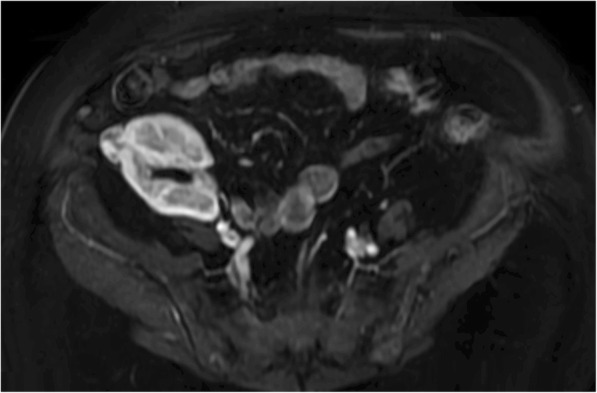
Fig. 1.

Magnetic resonance imaging (MRI) was used for the follow-up evaluation of renal mass because of its multi-planar capabilities, as well as its ability to demonstrate of enhancement and to provide soft-tissue contrast. A MRI scan at 60 months after transplantation showed a well-circumscribed oncocytoma with expansive net margins at the cortical surrounding of the kidney (white arrows). It has not infiltrative margins with extension in peri-renal fat or renal sinus, in the medulla of the kidney or in the major renal venous vessels
