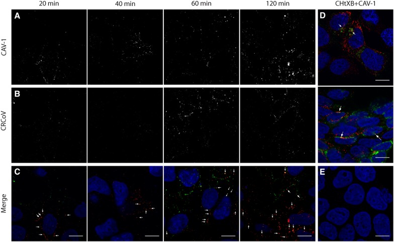
Figure 8.
CRCoV co-localize with caveolin. Cells treated with virus were synchronized on ice for 60 min and incubated at 37 °C for 20, 40, 60, or 120 min before they were washed and fixed. Caveolin-1 are presented on A (red channel) while B shows stained virus nucleocapsid protein (green channel). C Depicts merged image with marked co-localization. D FITC conjugated CHTxB (green) co-localizing with caveolin-1 (red) after 40 (upper) and 60 (lower) min. E Negative controls for staining. Scale bar 10 µm.

