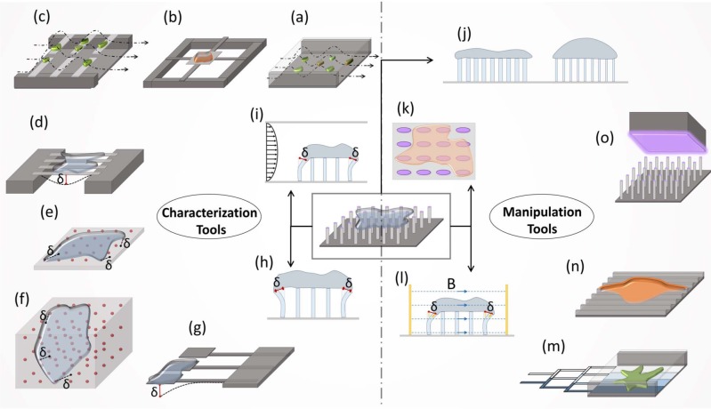
Figure 2.
BioMEMS devices in cell mechanics. The tools can be divided into two main categories: characterization tools, for the measurement of the different physical properties of cells, and manipulation tools, for the exertion of an extrinsic effect. (a) The adhesion strength characterization of cells in microfluidic channels is performed by simply counting the cells remaining after shear flow application. (b-c) Measurement of cell mass (b) in microfluidic chip and (c) on pedestals. Both tools are based on the resonance frequency change of the cantilevers or pad after cell attachment. (d) Cellular deformation measurement is performed by using piezoelectric nanoribbons. (e-i) The characterization of traction forces; (e-f) on 2D or in 3D bead embedded gels from the relative displacement of beads on (g) cantilever pads and (h) vertical micropillars is performed by measuring the deflection of cantilevers or micropillars, and (i) on micropillars under shear flow from micropillar displacement. (j-k) The manipulation of the cells by substrate alterations with micropillar configurations of (j) variable stiffness or (k) anisotropic pillar geometry. (l) Deformation application is performed using magnetic nanowires embedded in micropillars in a magnetic field. (m) The generation of substrate gradients is performed via microfluidics. (n) The manipulation of cell shape and phenotype is performed using nanoridge topography. (o) The generation of substrate patterns is performed using microcontact printing. Micropillar and microfluidic based approaches were found to have a variety of applications as both characterization and manipulation tools.

