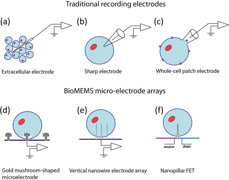
Figure 4.
Different forms of electrophysiological recording techniques. Shown above are schematic illustrations of traditional electrodeneuron interface configurations and BioMEMS microelectrode arrays. In the schematics, neurons are depicted in light blue (somas are marked with orange) (a) Extracellular recording electrode. The electrode does not penetrate any of the cells, thus it can record the activity of multiple neurons. (b) Intracellular recording with a sharp glass microelectrode. (c) Whole-cell patch clamp technique. This technique allows us to study single or multiple ion channels (marked with purple) located on a membrane patch of a single cell. (d) Gold mushroom-shaped microelectrodes are actively engulfed by neurons because of their dendritic spine-like shapes. The mushroom-shaped protrusion is 1.42μm high. (e) A vertical nanowire electrode array (VNEA) that penetrates the cell membrane providing direct contact with the cell. (f) A pillar-shaped protruding nanowire is the sensing gate electrode of the FET.

