Figure 2.

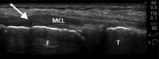
Ultrasound image of proximal MCL tear (arrow), noting ligament tissue disruption with retraction. Both the distal femur (F) and proximal tibia (T) are depicted with the adjoining MCL.

Ultrasound image of proximal MCL tear (arrow), noting ligament tissue disruption with retraction. Both the distal femur (F) and proximal tibia (T) are depicted with the adjoining MCL.