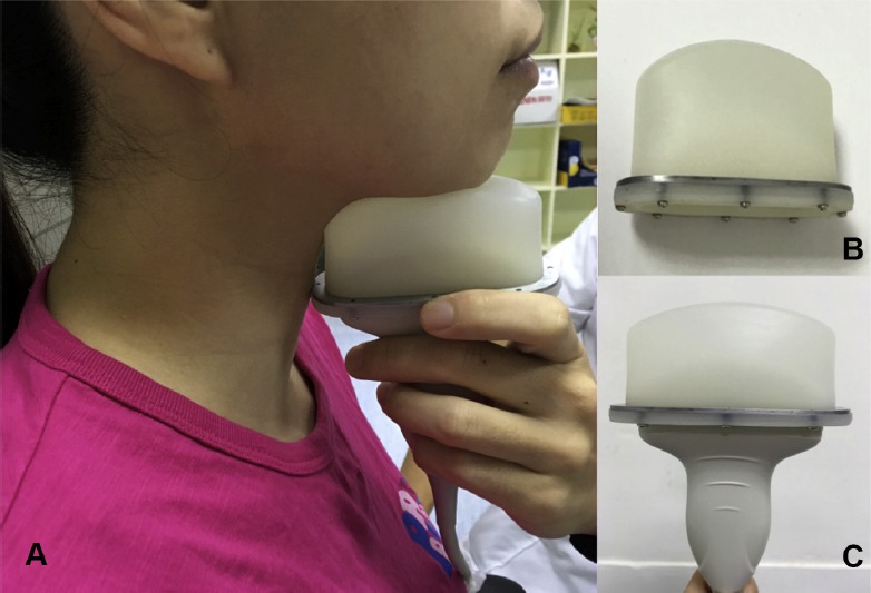
Figure 1.

Submental ultrasonographic examination. (A) The transducer is held in the vertical midsagittal plane at the mandible angle. (B) A self-designed water-filled probe cap, which enables tight and comfortable contact between the submental skin and the transducer. (C) The water balloon could be securely fixed onto the transducer.
