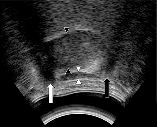
Figure 2.

Submental midsagittal ultrasonography image. It shows acoustic shadows behind the hyoid bone (black arrow) and the mandible (white arrow); between them are the suprahyoid muscles (between the white arrowheads). The tongue is above the suprahyoid muscles (between the black arrowheads).
