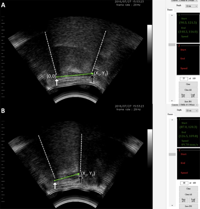
Figure 3.

Calculation of the hyoid bone displacement using a self-designed program. (A) The mandible (white arrow) and the hyoid bone (black arrow) were located at the intersections of the acoustic shadows (dashed lines) and suprahyoid muscles. Using a two-axis coordinate system and the mandible as the reference point, the position of the hyoid bone was designated a pair of coordinates (X1, Y1). (B) During swallowing, the recorded images were analyzed frame by frame to determine the maximal displacement of the hyoid bone from the resting position. The new position of the hyoid bone (black arrow) was designated as X2, Y2, with the mandible (white arrow) as the reference point. The distance between the two coordinates before and after swallowing denoted the hyoid bone displacement.
