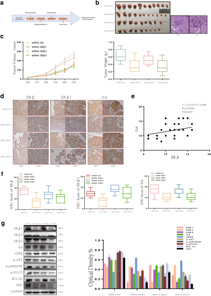
Fig. 6.
ERβ isoforms play differential roles in E2-stimulated IL6 expression. (a) The creation of our xenograft mouse model illustrated in a simplified sequence flow diagram. (b-c) Ovariectomized mice were subcutaneously injected with A549 cells transfected with shRNA-NC, shRNA-ERβ1, shRNA-ERβ2 or shRNA-ERβ5 shRNA lentiviral particles (GeneChem) (N = 8/group). Mice were euthanized and photographed after 4 weeks of E2 (0.09 mg/kg) treatment. The tumor photograph, tumor weight and tumor growth curves of each group were obtained. (d-f) IHC staining of ERβ, ERβ1 and IL6 showed a significant positive linear correlation between ERβ and IL6 expression. All data are presented as the mean of three independent experiments ± SD. (g) The protein expression of ERβ subtypes, IL6, p-p38MAPK, p-AKT and p-Stat3 in murine lung tumors was measured using western blot. All data are expressed as the mean ± SD. Student’s t-test was performed to assess statistical significance

