Figure 2.

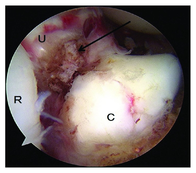
Case 1: arthroscopic photograph shows the fracture site (arrow). R: radial head; U: ulna (fracture bed); C: coronoid process.

Case 1: arthroscopic photograph shows the fracture site (arrow). R: radial head; U: ulna (fracture bed); C: coronoid process.