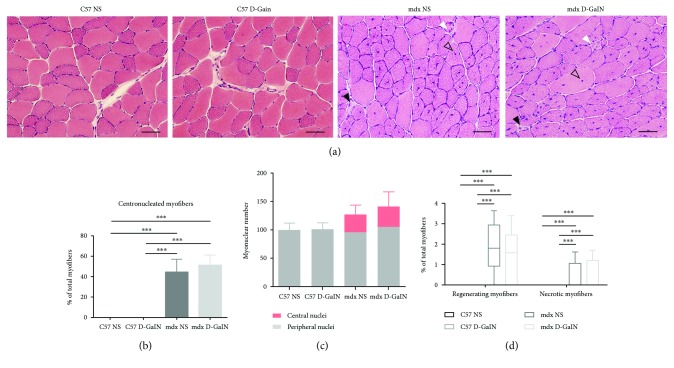
Figure 1.
Necrosis and regeneration in the muscle of D-galactosamine-treated and D-galactosamine-untreated mice. Mice were intraperitoneally injected with D-galactosamine (D-GalN) or normal saline (NS), and tibialis anterior (TA) was sectioned for histological analysis 24 h later. (a) HE staining showed normal morphology of TA in both the C57 NS and the C57 D-GalN groups and muscle lesions in both the mdx NS and the mdx D-GalN groups. Solid black arrows: necrotic myofibers with pale and homogenous sarcoplasm and pyknotic nuclei; solid white arrows: regenerating myofibers with a small diameter and vesicular central nuclei; and hollow black arrows: centronucleated myofibers. Scale bar: 100 μm. (b, c) The percentage of centronucleated myofibers in total myofibers (b) and the ratio of central versus peripheral nuclei (c) of different groups. (d) The percentage of regenerating and necrotic myofibers in total myofibers of different groups. ∗∗∗P < 0.001.

