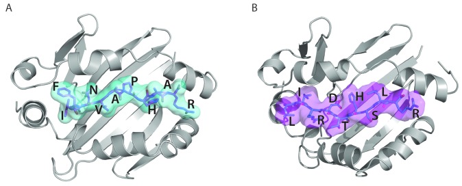
Figure 5.
Presentation of ADAMTS13 peptides on MHC class II. (A) Three-dimensional model structure of HLA-DRB1*11 (graphic representation in gray) with FINVAPHAR peptide (stick representation in magenta with cyan surface) from ADAMTS13 CUB2 domain. (B) Three-dimensional model structure of HLA-DQB1*03 (graphic representation in gray) with LIRDTHSLR peptide (stick representation in cyan with magenta surface) from ADAMTS13 CUB2 domain.

