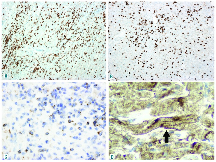
Figure 2.
Characterisation of lymphocytic infiltrates in the myocardium. Dense cytotoxic T-cell infiltrates were observed within the myocardium and the skeletal muscle. Representative image of immunostaining against CD8+ (A, 100×), granzyme B (B, 100×) and perforin (C, 100×) in myocardium slides. Cardiomyocytes (arrow) around necrotic areas showed intense membrane PD-L1 expression (D, PD-L1 clone 22C3, 100×).

