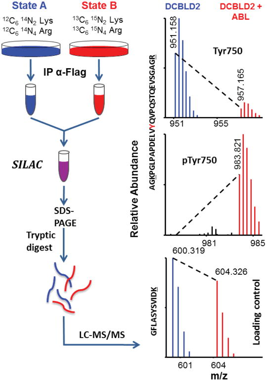
Figure 4.
Schematic of sample preparation for SILAC quantification and representative example of quantification by LC-MS/MS. Cells grown in media supplemented with either light or heavy lysine and arginine were transfected with DCBLD(X)–Flag in kinase inactive/inhibited (light) or active (heavy) conditions. Post-IP, heavy and light immune complex pairs were combined and analyzed via LC-MS/MS to determine relative abundance of phosphopeptide ions between each state. Heavy-to-light ratios (H:L) of monoisotopic peak intensities were normalized to DCBLD(X) peptides that were found not to be modified (loading control).

