Fig. 3.
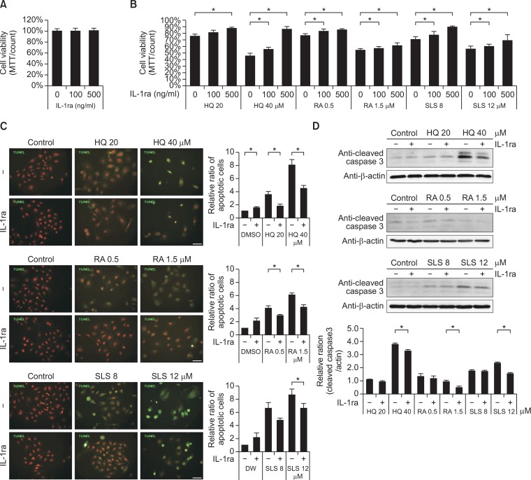
Recombinant IL-1ra treatment reduced cell apoptosis, which was induced by the chemical irritation. (A, B) MTT assay in keratinocytes treated without (A) or with HQ, RA, or SLS (B) of the same doses as in Figure 1 in the presence of absence of different doses of rhIL-1ra. Data in the graph represents mean ± SD of relative values compared to solvent-treated control from 3 to 5 independent experiments (*p<0.05 vs. solvent-treated control of corresponding chemical concentration). (C) TUNEL assay using Apo-BrdU TUNEL assay kit (green fluorescence) and (D) Western blot analysis of cleaved csapase-3 in keratinocytes treated with HQ, RA, or SLS using the same doses as in Fig. 1 in the presence of absence of 500 ng/ml of rhIL-1ra. Nuclei were counter-stained with Hoechst 33258 (scale bar=50 μm). Data in the graph represents mean ± SD of relative values compared to solvent-treated control from 3 independent experiments (*p<0.05 vs. corresponding chemical concentration without rhIL-1ra treatment).

