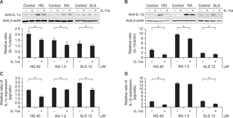
Fig. 4.
Recombinant IL-1ra reduced chemical irritation-induced IL-1α and IL-1β expression. Western blot analysis of (A) IL-1α and (B) IL-1β in cell lysates and ELISA of (C) IL-1α and (D) IL-1β in culture supernatants at 48 hrs after treatment with HQ, RA, or SLS using the same doses as in Fig. 1. Data in the graph represents mean ± SD of relative values compared to solvent-treated control from 3 independent experiments (*p<0.05 vs. corresponding chemical concentration without rhIL-1ra treatment).

