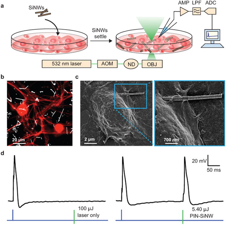
Figure 3. Basic silicon nanowire-based neural interfaces.

(a) Schematic of the set-up used to record DRG neuron APs in response to a 532 nm laser stimulation at a single neuron/SiNW interface. This set-up includes an ordinary electrophysiology capability for patch clamp experiments, with an amplifier (AMP), used in current clamp mode, a low pass filter (LPF), and an analog to digital converter (ADC). In addition the setup was implemented with a 532 nm laser beam controlled by an acoustic modulator (AOM) and various neutral density (ND) filters (to attenuate the power). The laser beam is aligned to the optical central axis of the objective lens (OBJ) of an inverted microscope. SiNWs were sonicated off of their growth substrate and drop casted onto the primary neuron culture. After 20 min of settling, the experiments were performed. (b) Confocal microscopy image of primary neonatal rat dorsal root ganglion (DRG) neurons stained with anti-β-tubulin III (red) co-cultured with PIN-SiNWs (white). This is a representative image from one of a total of 10 images taken from 2 independent experiments. (c) Scanning electron microscopy images of a single DRG neuron interacting with a single PIN-SiNW (left); zoomed in image displaying neuron-PIN-SiNW interface (right). This is a representative image from one of a total of 63 images taken from 4 independent experiments. (d) Patch clamp electrophysiology current clamp trace of membrane voltage in DRG neuron stimulated (blue pulse) or a laser pulse (green bar) of various energies as labeled at the neuron/SiNW interface. Two conditions are displayed: laser only with no SiNW (left) and PIN-SiNW (right). These are representative traces from one of 173 total action potential traces measured from 30 neurons with PIN-SiNWs, and one of 27 total traces from 2 neurons with PIN-SiNWs.
