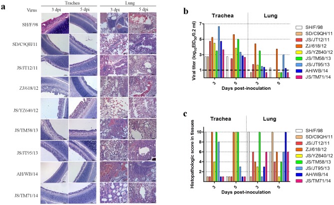
Fig 7. Histopathological changes and virus titers in lungs and tracheae of infected SPF chickens.
a) Histopathology of representative infected SPF chickens. Lungs and tracheae were collected at 4 dpi and fixed in 10% formalin, embedded in paraffin, and sectioned. b) Infection of the isolates in chickens. We inoculated groups of chickens intranasally and via the conjunctiva with 106 EID50 of the virus. Tracheae and lungs were harvested on 3 and 5 dpi. Viral titers for the tracheal and lung homogenates were determined by endpoint titration in SPF-embryonated chicken eggs. Each patterned bar represents the viral titers from an individual chicken. The black-dashed horizontal line indicates the lower limit of detection. c) Tissue sections were inspected, and histopathological changes were scored as follows. For the tracheae, 0: normal; 1: congestion; 2: cilia loss; 3: little inflammatory cell infiltration; and 7: a lot of inflammatory cell infiltration. For the lungs, 0: normal; 1: congestion; 2: hemorrhage; 3: inflammatory cell infiltration in the bronchial submucosa; and 7: a lot of inflammatory cell infiltration in the bronchial submucosa and alveolus. Average values for three birds are shown. Data are representative of three independent experiments.

