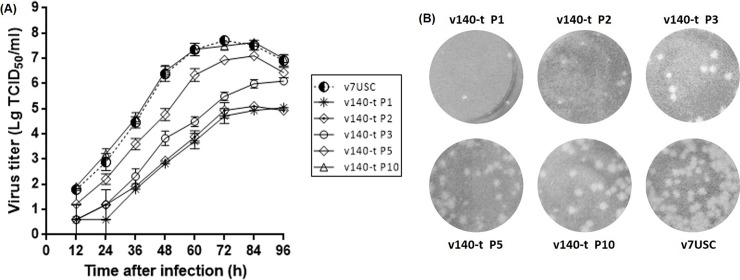
Fig 7. Viral growth and plaques characteristics of v140-t.
A: Analysis of v140-t (P1, P3, P5 and P10) growth in MARC-145 cells. Culture supernatants were collected at the indicated times and titrated. B: Analysis v140-t (P1, P3, P5 and P10) plaques characteristics in MARC-145 cells. MARC-145 monolayers were infected with v7USC, v140-t P1, v140-t P3, v140-t P5, and v140-t P10, respectively, overlaid with low melting-point agarose and stained with 5% (w/v) crystal violet in 20% ethanol at 5 dpi.

