Figure 4.

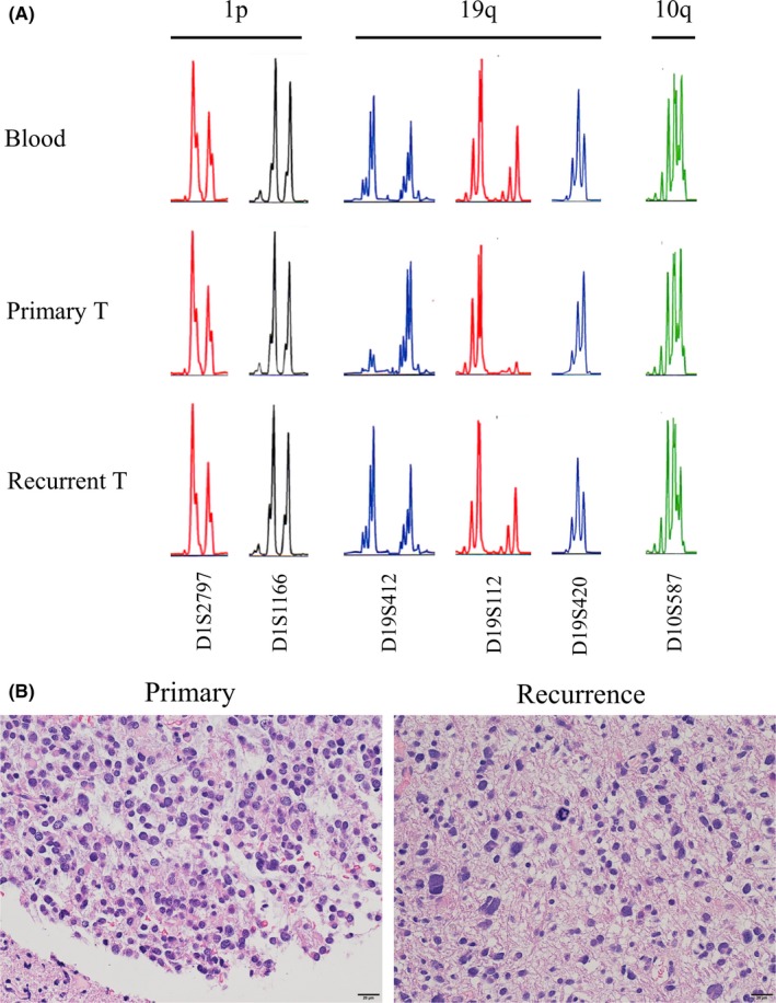
Alteration of the 19q status and histology between the primary and recurrent tumors (T) in case 3, a 40‐y‐old man with a left frontal glioma. A, Microsatellite analysis showing 1p/19q/10q loss of heterozygosity status of primary tumor and recurrent tumor compared with the constitutional DNA. Results of representative primers are shown. Primary tumor showed 1p‐intact, 19q‐loss, and 10q‐intact. Recurrent tumor showed 1p/19q‐intact, losing the19q‐loss status, and also showed new 10q‐loss. B, H&E staining (scale bar, 20 μm). Change in histological features between the primary and recurrent tumors is shown. Oligodendroglioma‐like cells observed in the primary tumor disappeared after recurrence
