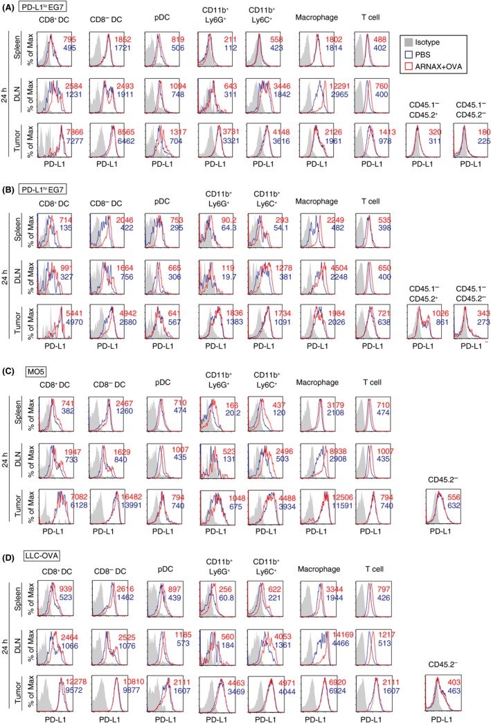
Figure 3.

Programmed death ligand‐1 (PD‐L1) expression on intratumor myeloid cells is different in each tumor type. A‐D, PD‐L1lo EG7 and PD‐L1hi EG7‐bearing CD45.1 congenic mice (C57BL/6) and MO5‐ and LLC‐ovalbumin (OVA)‐bearing WT mice (CD45.2) were treated with PBS or 10 μg ARNAX‐140 + OVA on day 7 (PD‐L1lo or hi EG7‐bearing mice) or day 10 (MO5‐ and LLC‐OVA‐bearing mice). After 24 h, spleens, draining lymph nodes (DLN) and tumor tissues were harvested, and cell suspensions were mixed in each group. PD‐L1 expression levels on immune cells in spleen, DLN and tumor were evaluated. In the case of PD‐L1lo or hi EG7 tumors, tumor cells (CD45.2) and host immune cells (CD45.1) were distinguished using CD45.1 and CD45.2 markers. PD‐L1 expression on intratumor non‐immune cells (the CD45.1−CD45.2+ population in EG7 tumors = tumor cells; the CD45.1−CD45.2− population in EG7 tumors = mesenchymal cells; and the CD45.2− population in MO5 and LLC‐OVA tumors = tumor cells and mesenchymal cells) were also analyzed. Results in PD‐L1lo EG7‐ (A), PD‐L1hi EG7‐ (B), MO5‐ (C) and LLC‐OVA‐bearing mice (D) are shown. Numbers in the histogram indicate mean fluorescence intensity
