
Figure 4.
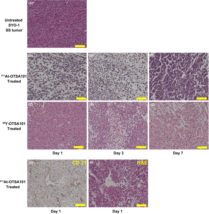
Histopathological analyses of the synovial sarcoma (SS) xenografts by immunohistochemistry and H&E staining. A, Untreated SYO‐1 SS xenograft. B,C, Day 1, D,E, day 3, F,G, day 7 after radioimmunotherapy. B,D,F, 211At‐OTSA101. C,E,G, 90Y‐OTSA101. H,I, CD31 immunostaining and its corresponding H&E staining at day 1 after 211At‐OTSA101 treatment. Scale bars, 50 μm