Figure 1.

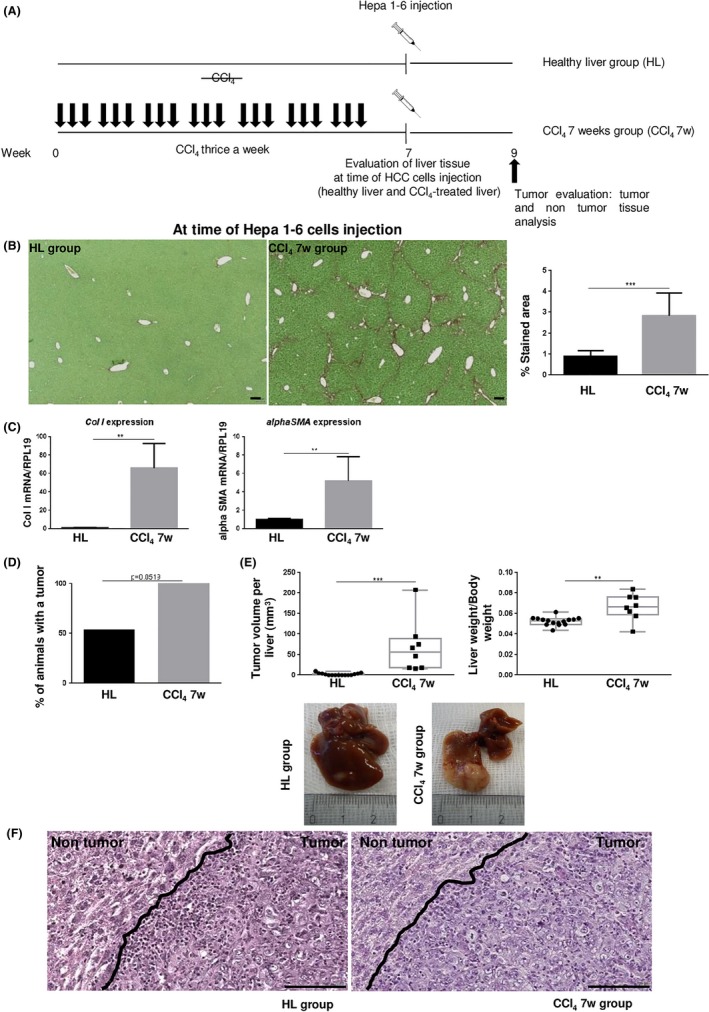
A, Comparison of hepatocellular carcinoma (HCC) cell growth when Hepa 1‐6 cells were injected into a healthy liver (HL group) and in mice pretreated with carbon tetrachloride (CCl4) for 7 wk (CCl4 7w group). B, Sirius red‐stained liver sections in CCl4‐treated (at time of HCC cell injection) and non‐treated mice. Scale bar, 100 μm. Collagen fibers were evaluated as percentage of stained area in the section (mean ± SD). C, Hepatic gene expression of Collagen I (Col I) and alphaSma (mean ± SD) at time of HCC cell injection. D,E, Percentage of animals with a tumor was calculated in the HL and CCl4 7w groups and tumor burden was evaluated by total tumor volume per liver (mm3) and the liver to body weight ratio. F, Representative H&E‐stained tumor and non‐tumor tissue sections. Scale bar, 100 μm. **P < .01; ***P < .001
