Figure 1.

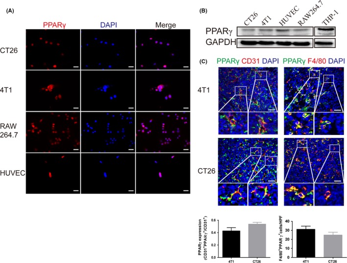
Peroxisome proliferator‐activated receptor γ (PPARγ) is expressed in tumor cells, endothelial cells and tumor‐associated macrophages (TAM). A, Immunofluorescence staining for PPARγ expression in cultured murine colon carcinoma cell line CT26, breast cancer cell line 4T1, macrophage line RAW264.7 and HUVEC. Scale bar, 20 μm. B, Western blot analysis of PPARγ expression in lysates from cultured 4T1 cells, CT26 cells, HUVEC and RAW264.7 macrophages. THP‐1 cells as positive controls. C, Double immunofluorescence staining for CD31 and PPARγ shows PPARγ expression in the endothelium of s.c. transplanted 4T1 and CT26 tumors. Double immunofluorescence staining for F4/80 and PPARγ indicates PPARγ expression in TAM of s.c. transplanted 4T1 and CT26 tumors. Scale bar, 50 μm
