
Figure 2.
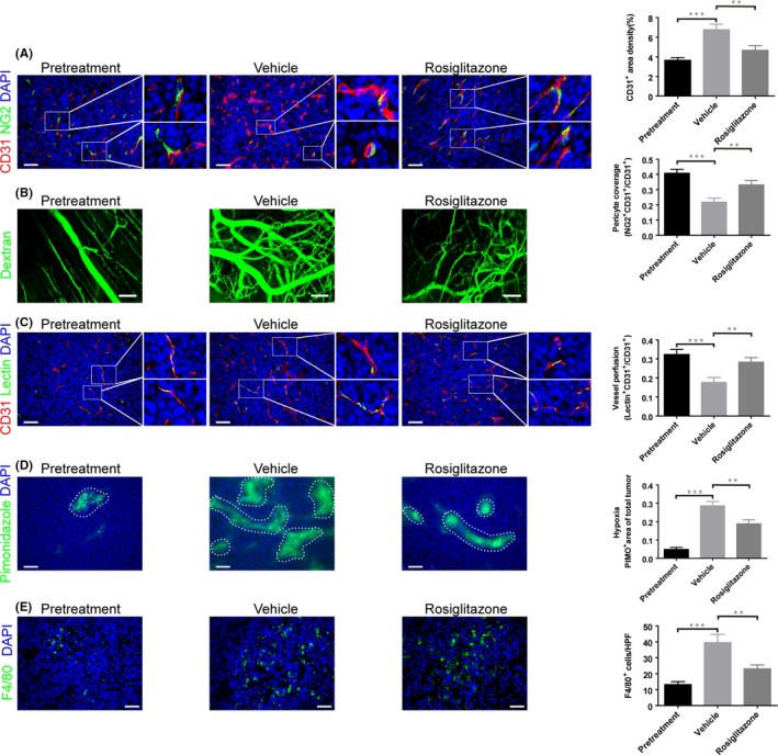
Rosiglitazone normalizes colon carcinoma vasculature and reduces tumor‐associated macrophage (TAM) infiltration. When CT26 tumor size approximately reached 50 mm3, the mice were given 100 mg/kg rosiglitazone or vehicle by oral gavage per day for 2 weeks. Then, the tumor samples were collected. A, Microvascular density and pericyte coverage were investigated by double immunofluorescence staining for CD31 and NG2. Scale bar, 50 μm. B, Multiphoton laser‐scanning microscopy images of CT26 tumor vasculature (green) stained with FITC‐dextran. Scale bar, 100 mm. C, Tumor vessel perfusion indicated by CD31‐positive endothelial cells stained red; FITC‐lectin perfused vessels are stained green. Scale bar, 50 μm. D, Representative micrographs show pimonidazole (PIMO)‐stained (green) CT26 tumor sections for hypoxia study. Scale bar, 100 μm. Hypoxic tumor regions are highlighted by dashed lines. E, Immunofluorescence staining for F4/80 showing TAM infiltration. Scale bar, 50 μm. Immunofluorescence images from (A to E) were captured within randomly selected fields (4‐6 fields per tumor, n = 6 mice). Data are presented mean ± SEM. **P < .01; ***P < .001