. 2018 Jul 3;7:e33910. doi: 10.7554/eLife.33910

Figure 1. DLK is necessary for the development of mechanical allodynia and induction of injury markers after spared nerve injury (SNI).

(A) DLK deletion is protective in the von Frey behavior assay following SNI at 1, 2, 3 and 4 weeks post injury (wpi). Baseline (BL) mechanical sensitivity thresholds are comparable between DLK WT (n = 9) and DLK cKO (n = 11). ****p<0.0001 by repeated measures ANOVA. (B) Scatter graph to illustrate individual points for DLK WT and DLK cKO at 4 weeks post injury. (C–H) Deletion of DLK reduces injury-induced transcription factors p-c-Jun and ATF3 in DRG following SNI. Representative images of ipsilateral L4 DRG 1 week after SNI stained for p-c-Jun in DLK WT (C) and DLK cKO (D) quantified in (E), and for ATF3 in DLK WT (F) and DLK cKO (G) quantified in (H). n = 6 per genotype. Scale bar in (G) valid for all images in this figure: 20 µm. ***p<0.001 by 2-tailed Student’s t test.

Figure 1—source data 1. data plotted in Figure 1A; von Frey behavior testing in DLK WT and cKO mice.
DOI: 10.7554/eLife.33910.006
Figure 1—source data 2. data plotted in Figure 1E; p-cJun-positive nuclear counts in ipsilateral L4 DRG 7 days after SNI in DLK WT vs cKO mice.
DOI: 10.7554/eLife.33910.007
Figure 1—source data 3. data plotted in Figure 1H; ATF3-positive nuclear counts in ipsilateral L4 DRG 7 days after SNI in DLK WT vs cKO mice.
DOI: 10.7554/eLife.33910.008
Figure 1—source data 4. data plotted in Figure 1—figure supplement 1; dynamic brush scores 7 weeks after SNI in DLK WT vs cKO mice.
DOI: 10.7554/eLife.33910.009
Figure 1—source data 5. data plotted in Figure 1—figure supplement 2; von Frey testing in DLK WT vs cKO mice at 1, 3, 5 and 7 days post SNI.
DOI: 10.7554/eLife.33910.010
Figure 1—source data 6. data plotted in Figure 1E; p-cJun-positive nuclear counts in ipsilateral L4 DRG 48 hr after hind paw formalin injection in DLK WT vs cKO mice.
DOI: 10.7554/eLife.33910.011
Figure 1—source data 7. data plotted in Figure 1E; ATF3-positive nuclear counts in ipsilateral L4 DRG 48 hr after hind paw formalin injection in DLK WT vs cKO mice.
DOI: 10.7554/eLife.33910.012

Figure 1.

Figure 1—figure supplement 1. DLK deletion prevents allodynia to dynamic mechanical stimulus after spared nerve injury (SNI).

Figure 1—figure supplement 1.

Dynamic brush testing at 7 weeks post injury showing that DLK cKO mice display less nocifensive and protective behaviors compared with DLK WT littermates (n = 6 per genotype group; 3 males and three females). Each data point represents the average of 4 trials per animal. Score scale: 0 – rapid paw lifting; 1 – paw guarding; 2 – strong paw lifting/kicking; 3 – paw licking. **p<0.01 by 2-tailed Student’s t test. ns: not significant.
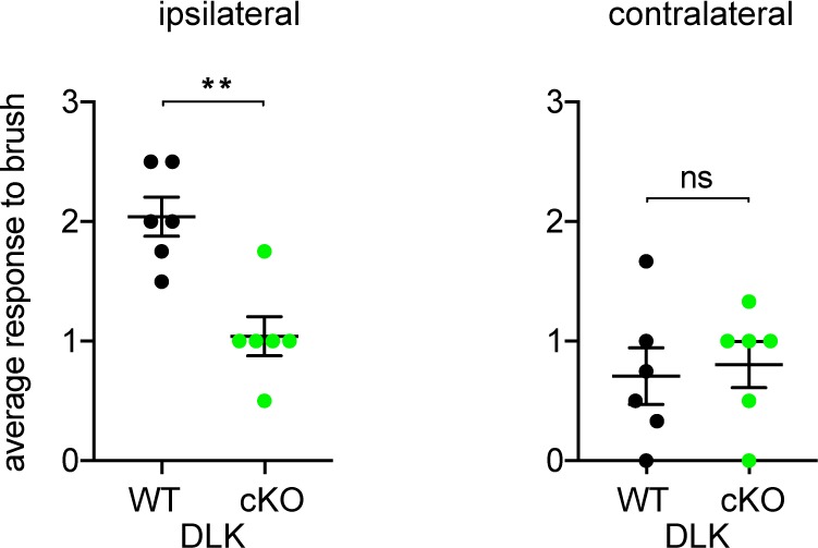
Figure 1—figure supplement 2. DLK is necessary for the development of mechanical allodynia after spared nerve injury (SNI).

Figure 1—figure supplement 2.

Von Frey testing at acute time points 1, 3, 5, and 7 days post injury (dpi) in DLK cKO (n = 11, 5 females and 6 males) and DLK WT littermates (n = 12, 6 per sex). DLK deletion is protective in the von Frey behavior assay following SNI at 1, 5, and 7 days post injury (dpi) by ordinary two-way ANOVA with filament size and genotype as factors. P values for a genotype effect were 0.0002 (1 dpi), 0.0038 (5 dpi), and p<0.0001 (7 dpi). Asterisks under green data points represent significant p values for differences between genotypes at individual filaments by Sidak’s multiple comparisons post test. *p<0.05, **p<0.01, ***p<0.001. The apparent hypersensitivity of the DLK WT group was reduced at 3 and 5 dpi, perhaps due to the aversive nature of paw withdrawal causing additional pain due to movement during this acute recovery period 1–5 days after injury.
Figure 1—figure supplement 3. DLK deletion reduces injury markers in DRG in the formalin model of neuropathic pain.

Figure 1—figure supplement 3.

(A–C) Example images of co-immunostaining for p-c-Jun (injury marker) and NeuN (all neuronal nuclei) in a wild type DRG to illustrate the % NeuN-positive neuron counts that were done for the data shown in Figure 1C–H and I–J in this figure. (D–J) Deletion of DLK reduces injury-induced transcription factors p-c-Jun and ATF3 in DRG following nerve injury by intraplantar formalin injection. Representative images of ipsilateral L4 DRG 48 hr after formalin treatment stained for p-c-Jun in DLK WT (D) and DLK cKO (E) quantified in (I), and for ATF3 in DLK WT (G) and DLK cKO (H) quantified in (J). (DLK WT n = 2; DLK cKO n = 3). (F) Representative image of p-cJun staining in a contralateral L4 DRG showing that p-cJun is normally low; compare with injury-induced p-cJun in D). Scale bar in (H) 100 µm, valid for all images in this figure except in (F) where scalebar = 50 µm). ***p<0.001 by two-tailed Student’s t test.