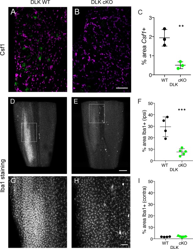
Figure 3. DLK is necessary for Csf1 upregulation in DRG neurons and for spinal cord microgliosis elicited by SNI.
(A–C) Representative images and quantification of Csf1 gene expression changes following SNI by in situ hybridization in DLK WT vs DLK cKO L3 DRG 7 days post SNI. Scale bar 100 µm. **p<0.01, by 2-tailed Student’s t test. Averaged data for n = 4–6 sections quantified in n = 3 mice per genotype. (D–I) Representative images and quantification of microgliosis in DLK WT vs DLK cKO spinal cords 8 days post-SNI. Iba1 staining in cleared spinal cords (top view, longitudinal plane) showing the SNI-induced microgliosis in the ipsilateral (left) dorsal horn of DLK WT spinal cord (D, G) that does not occur in the DLK cKO (E, H). (G) and (H) are higher magnification images of boxed areas in (D) and (E) respectively. Quantification of dorsal horn microgliosis in ipsilateral (F) and contralateral (I) spinal cord of DLK WT (n = 4) vs DLK cKO (n = 5). Scale bar in (D, E) 400 µm. Scale bar in (G, H) 100 µm. ***p=0.0010 by 2-tailed Student’s t test.

