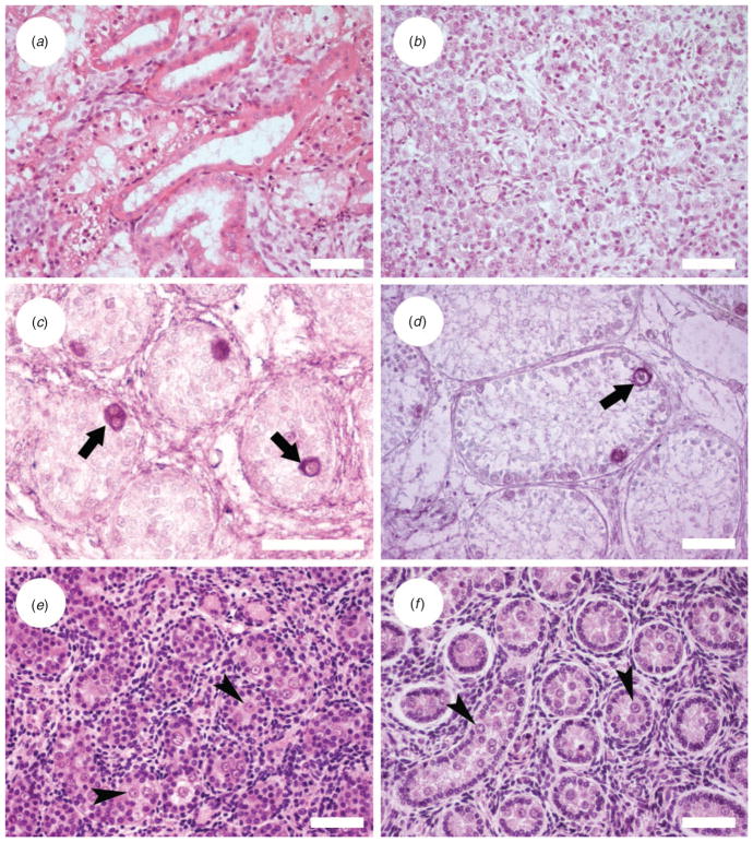
Fig. 1.
Histological appearance of donor testicular tissue. (a) Iberian lynx, 6-week-old fetus; (b) Iberian lynx, 1.5-day-old cub; (c) germ cells labelled by protein gene product (PGP) 9.5 immunostaining in a 6-month-old Iberian lynx testis tissue; (d) germ cells labelled by PGP 9.5 immunostaining in a 2-year-old Iberian lynx; (e) Cuvier’s gazelle aborted fetus; and ( f ) Mohor gazelle, 8-month-old male. Arrows indicate spermatogonia; arrowheads indicate gonocytes. Scale bars = 50 μm.

