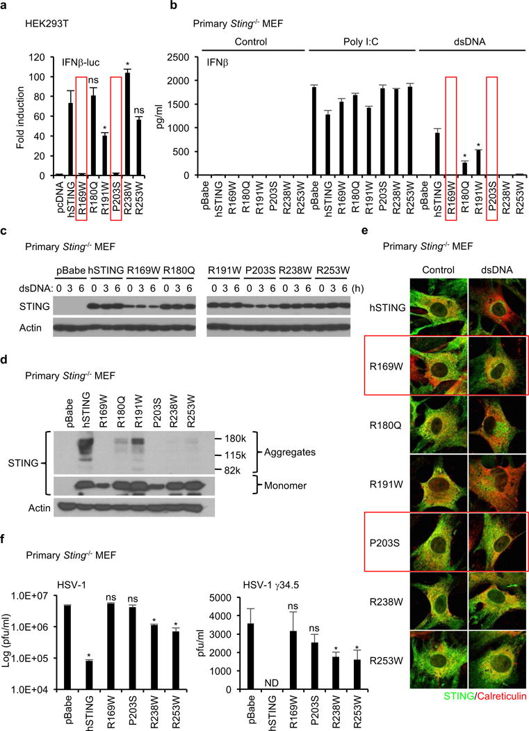
Figure 2.

STING mutants found in human tumors fail to activate dsDNA-induced inflammation. (a) The indicated plasmids were transfected into HEK293T cells with reporter plasmids (IFNβ-luc and TK-luc). After 24 hr, the luciferase activity in the cell lysates was measured. (b) Primary Sting−/− MEF cells were reconstituted with the indicated STING mutants using retrovirus. IFNβ in the supernatants was measured by ELISA after treated with Poly I:C (2 μg/ml) or dsDNA (4 μg/ml) for 16 hr. (c and d) The reconstituted Sting−/− MEF cells were treated with dsDNA (4 μg/ml) for the indicated times (c). Wester blots were performed with the indicated antibodies. (e) After treated with dsDNA as described in Figure 2c, the reconstituted Sting−/− MEF cells were stained with antibodies for STING and an ER marker, calreticulin, to observe the localization of STING using a confocal microscope. (f) The reconstituted Sting−/− MEF cells were infected with HSV-1 or HSV-1 γ34.5 for 24 hr and then the viral titer was determined by plaque assay. Data shown here are the averages ± SD (n = 3). Asterisks indicate significant difference (p < 0.05) compared to hSTING (a and b) or pBabe (f) determined by Student’s t-test (ns; not significant, ND; not detected).
