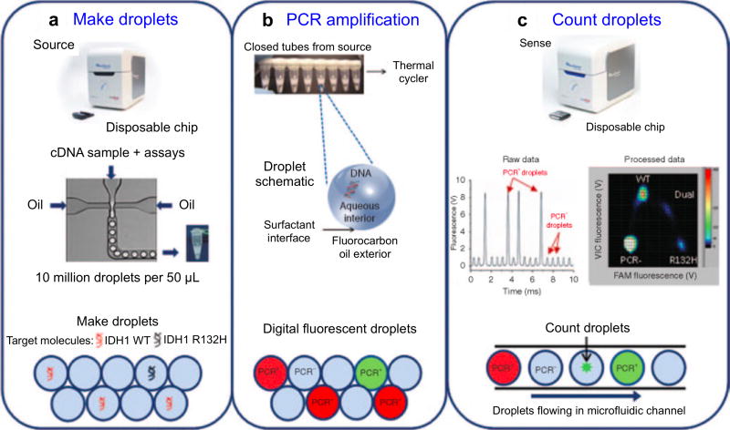
Figure 19. Application of droplet digital PCR for EV analyses.
The workflow consists of three steps: (a) Making PCR droplets. Aqueous PCR reaction mixture is injected through a microfluidic droplet generator along with surfactant-containing fluorocarbon oil to produce five picoliter droplets. (b) PCR amplification. The droplets are loaded into a standard thermal cycler for endpoint PCR amplification, with single-target-molecule-containing droplets resulting in specific probe hydrolysis (PCR+) and bright fluorescence and the majority of droplets, containing no target molecule, resulting in only background probe fluorescence (PCR−). (c) Each droplet's fluorescence is detected and processed into a two-dimensional scatter plot display. Reprinted with permission from Ref 94. Copyright 2013 American Society of Gene & Cell Therapy.

