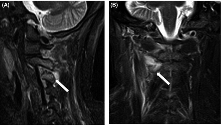
Figure 1.

A, Sagittal T2‐weighted fat saturation magnetic resonance imaging reveals erosion and a high‐intensity area around the C3‐C4 articular processes (white arrow). B, The coronal image reveals a high‐intensity area around the right C2‐C3 facet joints (white arrow)
