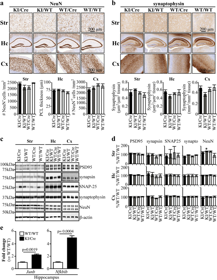
Fig. 4.
Differential synaptic profile mRNA expression in Casp6-expressing hippocampus, but not in striatum tissues. a, b Representative micrograph of 20–22-month-old KI/Cre, KI/WT, WT/Cre, or WT/WT hippocampus tissue sections immunostained with anti-NeuN (a) or anti-synaptophysin (b). Histograms represent the quantification of NeuN (a) and synaptophysin (b) immunostaining in the brain of 5 KI/Cre, 5 KI/WT, 5 WT/Cre, and 5 WT/WT mice. c, d Western blot and densitometric analyzes of synaptic and neuronal markers in the brains of aged (16–20 months) KI/Cre, KI/WT, WT/Cre, and WT/WT hippocampal protein extracts with antibodies against PSD-95, synapsin, SNAP-25, synaptophysin and NeuN. Quantitative results are represented as ratios of protein levels over β-actin and are arbitrarily expressed as percentage ± SEM of WT/WT. Statistical evaluations were done with ANOVA followed by a Dunnett’s post-hoc analysis against KI/Cre values. **p < 0.01, ***p < 0.001. e Differential synaptic plasticity profile mRNA expression of Junb and Nfkbib in KI/Cre versus WT/WT hippocampal tissues. No change was observed in striatum. Student’s t-test shows a significant difference between WT/WT and KI/Cre (n = 3 each)

