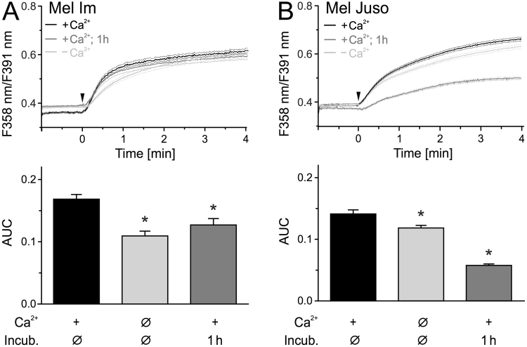
Figure 3.
Addition of CAP-pretreated solution onto cells leads to an immediate Ca2+ influx. Measurement of cytoplasmic Ca2+ using fura-2 AM. CAP exposed pbECS (100 µl) with Ca2+ was applied (black traces) onto Mel Im ((A), n = 485) and Mel Juso ((B), n = 321) 1 min after start of recording (arrowhead). The experiment was repeated with an interval of 1 h between CAP-exposure and application of the solutions (dark grey traces) and in the absence of extracellular Ca2+ (light grey trace) ((A), n = 274–411; (B), n = 366–579). Data are shown as mean and 99% confidence interval.

