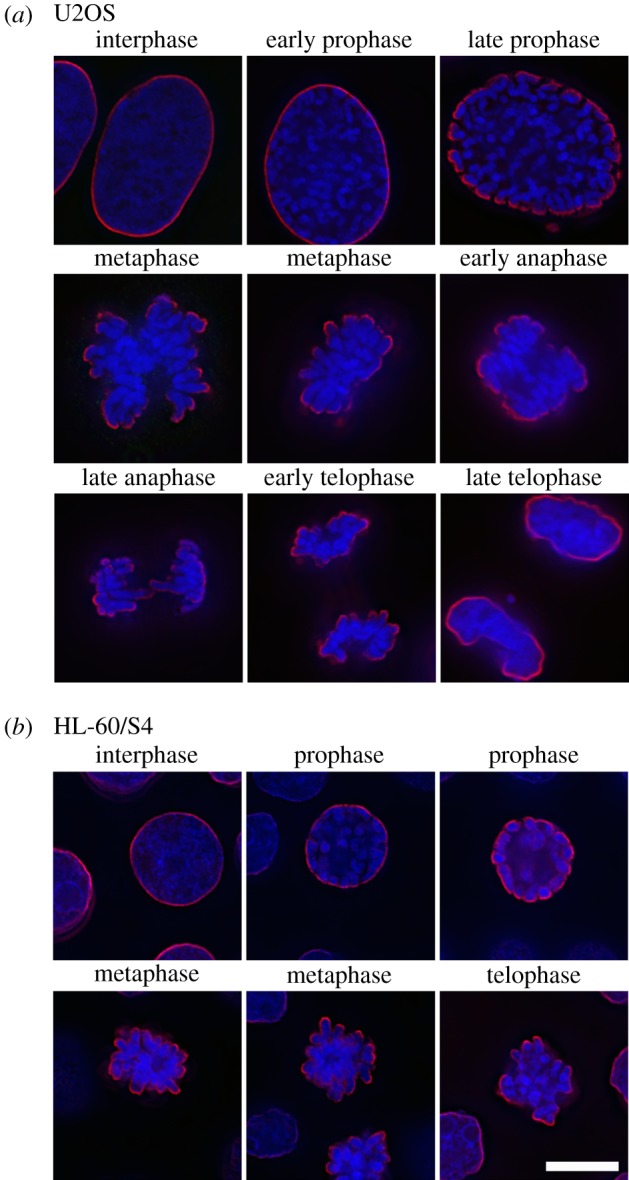
Figure 1.

Immunostaining of the epichromatin epitope throughout the cell cycle in U2OS (a) and HL-60/S4 (b) cells using mAb PL2-6 (red) and a DNA stain (DAPI, blue). Note that epichromatin staining persists on the outer edges of the mitotic chromosomes, even following NE breakdown. The magnification bar for (a) and (b) equals 10 µm. This image has been previously published [15].
