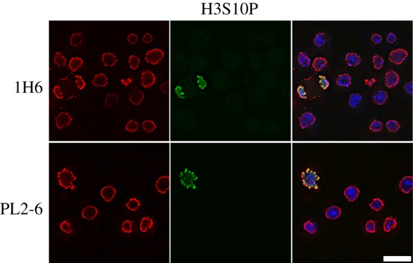
Figure 2.

Immunostaining of the epichromatin epitope in interphase and mitotic Drosophila Kc cells using two different mAbs that stain epichromatin (PL2-6 and 1H6, red), rabbit polyclonal anti-H3S10p (green) and DNA (DAPI, blue). Note the similar staining of epichromatin as shown for human U2OS and HL-60/S4 cells (figure 1). The magnification bar equals 10 µm. This image has been previously published [16].
