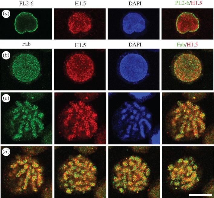
Figure 5.
Immunostaining patterns of bivalent PL2-6 and monovalent Fab fragments, derived from PL2-6. (a) An undifferentiated HL-60/S4 interphase nucleus stained with PL2-6 (green), anti-histone H1.5 (red), DAPI (blue) and merged red and green (R + G). (b) An undifferentiated HL-60/S4 interphase nucleus stained with Fab (green), anti-histone H1.5 (red), DAPI (blue) and merged (R + G). (c) A single confocal Z-slice from a mitotic HL-60/S4 cell stained with Fab, anti-histone H1.5, DAPI and the merged (R + G). (d) Various Z-slices from the merged mitotic R + G stack are presented. For all images, the magnification bar equals 10 µm. This image has been previously published in part [20].

