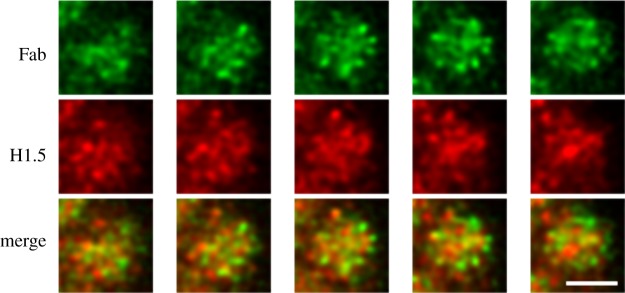
Figure 7.
Immunostaining patterns of monovalent Fab fragments on alternate sequential Z-slices along a mitotic chromosome arm, revealing a radial ‘chromomeric’ pattern. The top row is Fab (green); middle row, anti-histone H1.5 (red). The bottom row contains the merged ‘red + green (R + G)’ slices. Chromomeres are approximately 300 nm in diameter. The magnification bar equals 2 µm. This image has been previously published [20].

