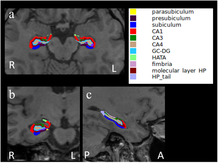
Figure 1.
The representative subdivision of the hippocampal subfields. The mask of each region overlapped on the axial (a), coronal (b), and sagittal (c) T1-weighted images. R, right; L, left; A, anterior; P, posterior. Color classification: parasubiculum, yellow; presubiculum, dark purple; subiculum, blue; CA1, red; CA3, dark green; CA4, brown; granule cell layer of dentate gyrus (GC-DG), sky blue; hippocampus-amygdala-transition-area (HATA), green; fimbria, purple; molecular layer hippocampus (HP), dark brown; hippocampal tail, gray.

