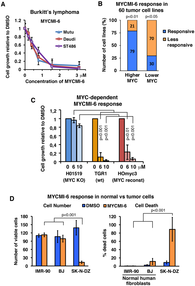
Figure 6.
MYCMI-6 inhibits tumor cell growth and viability in a MYC-dependent manner but is not cytotoxic to primary normal human cells. (A) MYCMI-6 titration on Burkitt’s lymphoma (BL) cell lines Mutu, Daudi and ST486. Data are shown as mean ± standard deviation of 2 biological experiments, each with 3 technical repeats. (B) Correlation MYCMI-6 response (GI50) with MYC mRNA levels of the NCI-60 human tumor cell lines extracted from CellMiner™ and complemented with MYC protein levels from the Novartis proteome scout project or from the literature (see Supplementary Table S1). “Responsive” and “less responsive”; cell lines with positive and negative log 10 GI50 values, respectively. “Higher MYC” and “lower MYC”; cell lines with higher and lower MYC expression levels (MYC mRNA/protein) than average, respectively. p-values are indicated. (C) Growth of TGR-1 (wt), HO15.19 (MYC knockout) and HOmyc3 (MYC reconstituted HO15.19) Rat1 fibroblasts, as measured by the WST-1 assay after treatment with MYCMI-6. Data are shown as mean ± standard deviation of 3–5 biological experiments, each with 3 technical repeats. p-values are indicated. (D) Normal IMR-90 and BJ human fibroblasts and the MYCN-amplified neuroblastoma cell line SK-N-DZ were treated with 12.5 μM MYCMI-6 or control (DMSO) for 24 hours. The number of viable and percentage of dead cells were quantified by addition of CellTracker Green (stains all cells) and DAPI (stains dead cells) to cells and analyzed in GFP and CFP channels using a fluorescence microscope. p-values are indicated.

