Figure 7.
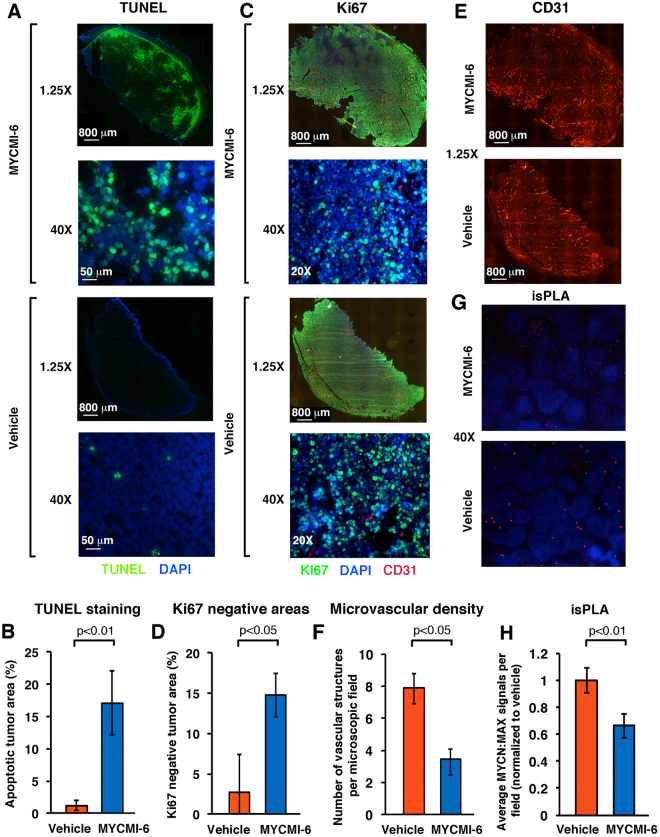
MYCMI-6 inhibits MYC:MAX interaction, induces apoptosis and reduces tumor cell proliferation and microvascularity in a MYCN-amplified neuroblastoma mouse tumor model in vivo. SK-N-DZ MYCN-amplified neuroblastoma xenograft tumors reaching a volume of 100–200 mm3 were treated with MYCMI-6 (20 mg/kg body weight) or vehicle injected i.p. daily for 1–2 weeks. (A) Apoptosis was determined by TUNEL staining (green) of tumor tissues from mice treated with MYCMI-6 (upper two panels) or vehicle (lower two panels), counterstained with DAPI (blue). Representative images are shown at 1.25X (panel 1 and 3 from top, bar = 800 μM) or 40X (panel 2 and 4, bar = 50 μM) magnification. (B) Quantification of TUNEL staining normalized to whole tumor areas as determined by DAPI from three MYCMI-6- and three vehicle-treated mice. (C) Cell proliferation and microvascular density determined by Ki67 (green) and CD31 (red) staining, respectively, of tumor tissues from mice treated with MYCMI-6 (upper two panels) or vehicle (lower two panels), respectively, and counterstained with DAPI (blue). Representative images taken at 1.25X (panel 1 and 3 from top, bar = 800 μM) or 20X (panel 2 and 4) magnification. (D) Quantification of Ki67 negative areas normalized to whole tumor areas by DAPI from three MYCMI-6- and three vehicle-treated mice. (E) microvascular density visualized by CD31 staining in the red channel at 1.25X magnification as in (C). (F) Quantification of CD31 staining normalized to whole tumor areas from three MYCMI-6- and three vehicle-treated mice. (G) Detection of MYCN:MAX protein interaction by isPLA performed on tumor tissue from mice treated with MYCMI-6 (upper panel) or vehicle (middle panel) using antibodies against MYCN and MAX. Representative images were taken at 40X magnification. (H) Quantification of MYCN:MAX isPLA signals in tumor tissue from MYCMI-6- and vehicle-treated mice, presented as average number of dots from four randomly chosen microscopic fields from MYCMI-6 treated mice normalized to corresponding values from vehicle-treated mice. SEM and p-values are indicated.

