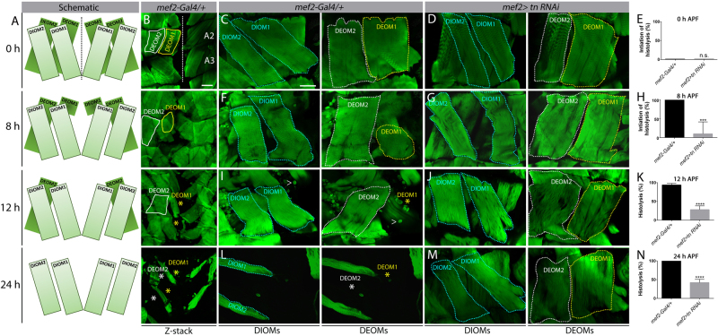
Fig. 1. Tn is required for DEOM histolysis.
a Schematic diagrams of the DEOMs during pupal development at 0, 8, 12, and 24 h APF. Dotted line denotes the midline. b Merged Z-stack images of DEOM histolysis that correspond to the same time points (a) in mef2-Gal4/+ control muscles stained to visualize F-actin (green). DEOM1 (yellow solid line) and DEOM2 (white solid line) are both present at 0 h APF. In WT muscles, DEOM1 starts to disintegrate at 8 h APF and is gone by 12 h APF (yellow asterisk). DEOM2 disappears by 24 h APF (white asterisk). A2 and A3 denotes abdominal segments 2 and 3, respectively. c–n Representative images and quantification of DEOM muscle histolysis at 0, 8, 12, and 24 h APF in mef2-Gal4/+ control or mef2>tn RNAi muscles stained with phalloidin (green). Substacks of single confocal planes separate out the DIOMs (cyan dotted lines) from the DEOMs. c, f, i, l DEOM1 (yellow dotted lines) and DEOM2 (white dotted lines) muscles degenerate (asterisks) by 24 h in control muscles. d, g, j, m However, reduction of tn by RNAi mostly blocks DEOM histolysis. e, h, k, n DEOM1 histolysis is not initiated at 0 h in mef2-Gal4/+ control or mef2>tn RNAi muscles (e). By 8 h, all control DEOM1s have started to breakdown, while most of these muscles are still present in tn RNAi pupae (h). By 12 h (k) or 24 h (n), most DEOM1s are still intact in tn RNAi muscles. White carets indicate remnants of fat body tissue that stain positive for F-actin. Mean ± SEM (n.s., not significant, ****p < 0.001, ***p < 0.005). Scale bars, 100 µm (b); 50 µm (c, d, f, g, i, j, l, m)

