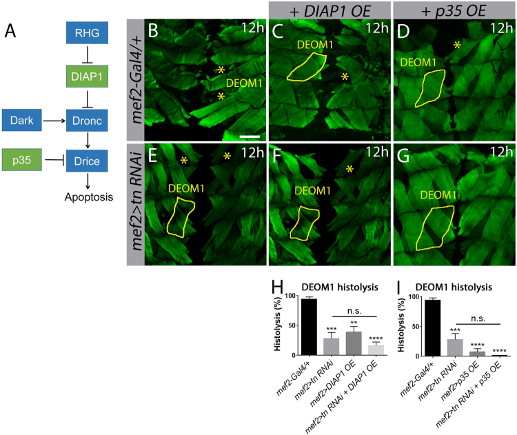
Fig. 4. tn genetically interacts with DIAP1 during DEOM histolysis.
a Schematic of Drosophila core cell death machinery. b–g Merged Z-stack confocal images of the abdominal muscles stained with phalloidin (green) at 12 h APF. DEOM1 is outlined with a solid yellow line and histolysed DEOM1s are marked by yellow asterisks. b Histolysis proceeds normally in mef2-Gal4/+ muscles. Overexpression of DIAPI (c) or p35 (d) in muscles partially blocks DEOM1 histolysis. e–g There is no significant difference in DEOM1 histolysis upon RNAi knockdown of tn alone (e), or with overexpression of DIAP1 (f) or p35 (g) in a tn RNAi background. h A bar graph showing similar levels of muscle histolysis in mef2>tn RNAi+DIAP1 OE compared to mef2>tn RNAi muscles. i Quantification showing no significant difference in DEOM1 breakdown between mef2>tn RNAi and mef2>tn RNAi+p35 OE pupae. Mean ± SEM (n.s., not significant, ****p < 0.001, ***p < 0.005, **p < 0.01). Scale bar, 100 µm (b–g)

