Figure 3.
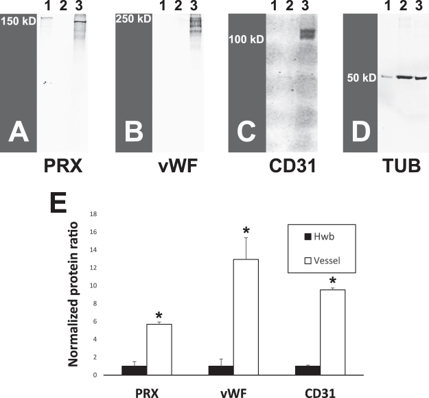
Expression of PRX protein in human brain vessel fractions. Fractions of human brain from autopsy were analyzed for presence of indicated proteins. Lane 1: 293 cells transfected with L-PRX full length cDNA (encoding Genbank Protein ID AAH67266.1; purchased from OriGene); Lane 2: Human whole brain (Hwb) lysate; Lane 3: Partially purified microvessel proteins from human brain. Lane 1 was underloaded to avoid overloading PRX. (A) Western blotting of brain vessels showed expression of a protein band corresponding to the larger form of PRX (L-PRX), as seen in transfected 293 cells. As controls, the same protein lysates were probed for (B) vWF, (C) CD31, and (D) tubulin. Quantification of the degree of enrichment of vascular proteins in vascular fractions was computed by first calculating the ratio of the protein of interest to tubulin (D) and then normalizing this ratio in vessels to the ratio in human whole brain. (E) shows overall levels of protein enrichment in vessel fractions of multiple individuals (n = 3 for PRX and vWF and n = 2 for CD31; *indicates p < 0.01). Additional brain samples and higher exposure PRX blots are shown in Supplemental Fig. 1.

