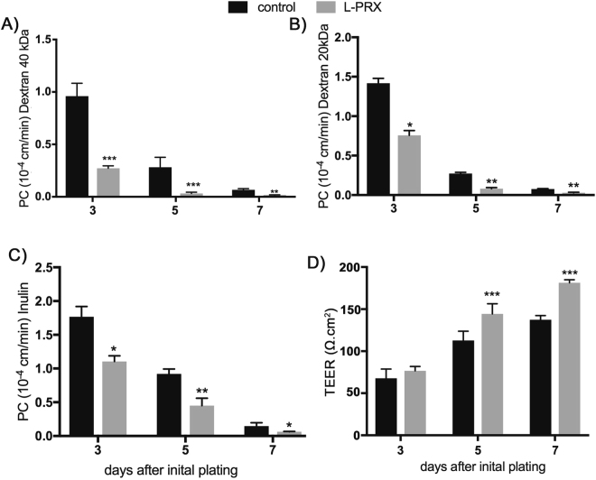
Figure 6.
Effect of PRX on mouse cerebral endothelial cell permeability. L-PRX was expressed in primary mouse cerebral endothelial cells using recombinant lentiviruses. Uninfected cells were used as a control. At the number of days after infection indicated on the x-axis, permeability to indicated fluorescent tracers was determined (A–C). (D) TEER was also determined at the same time points. L-PRX significantly strengthened the barrier function of endothelial cells, accelerating both the acquisition of barrier function and the final resistance of cells to tracers of a wide range of molecules. RFP lentivirus infected cells showed the same permeability profiles as uninfected cells (not shown). Data represent means +/− SD for n = 5 independent experiments. *p < 0.05, **p < 0.01, ***p < 0.001 comparing control and L-PRX transfected cells.

