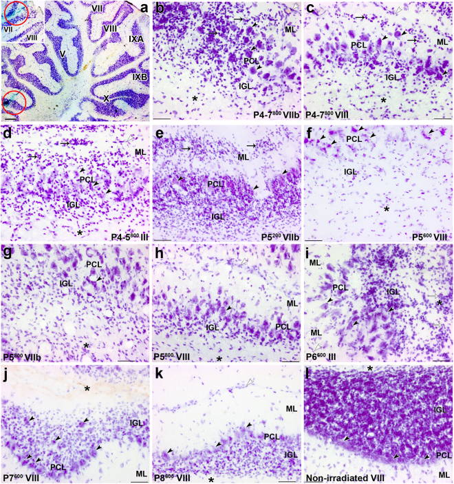
Figure 3.
Abnormal histological features in the cerebellar cortex of adult rats, induced by postnatal X-irradiation. (a) The tracks of the recording microelectrode can be reconstructed from the position of the two deposits of Fast green (red circles visible in lobules III (a) and VII (inset). (b–i) Abnormal arrangement of PCs (black arrowheads) in multiple layers (PCL) in lobules VIIb (b) and VIII (c) in Groups P4-7800, lobules III (d) in Group P4-5800, lobules VIIb (g) and VIII (h) in Group P5800 and lobule III (i) in Group P6600. PCs are more or less normally distributed in a monolayer in lobule VIII in Groups P7600 (j), P8600 (k) as well as in the non-irradiated control cerebellum (l). Ectopic granule cells (arrows) are located in the molecular layer (ML) and PCL in lobules VIIb (b) and VIII (c) in Groups P4-7800 and P5200 (e) and in lobule III (d) in Group P4-5800. In the atrophied internal granular layer (IGL), granule cells are extremely depleted in Groups P4-7800 (b,c), P4-5800 (d), P5800 (g,h), P5600 (f) and P6600 (i). These cells are partly restored in the IGL in Groups P7600 (j) and P8600 (k). Abnormal location of granule cells (white arrows) in the white matter (*) is seen in the lobule III in Group P6600 (i). White arrowheads indicate the surface of the cerebellar cortex in (b,c,d,h,i,k). Scale bars = 500 µm in a, 50 µm in b-l.

