
Figure 5.
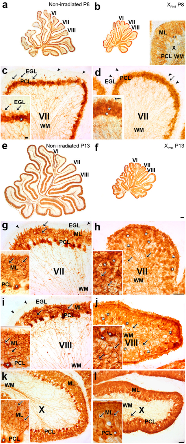
CaBP–immunohistochemical staining of Purkinje cells in the developing cerebellum of normal and postnatally X-irradiated rats from Group P5600. (a,b,e,f) Low magnification of sagittal sections of the cerebellar vermis of control (a,e) and X-irradiated Group P5600 (b,f) rats at P8 (a,b) and P13 (e,f). Insets b, c, d, g-l, High magnifications of sagittal sections of the late-developing lobules VII (c,d,g,h) and VIII (i,j) to compare with early-developing lobule X (b,k,l) in the cerebellar vermis of normal rats at P8 (c) and P13 (g,i,k) and Group P5600 X-irradiated rats at P8 (b,d) and P13 (h,j,l). PCs (white asterisks) are already disorganized in the 8-day-old X-irradiated lobule VII in (d). Short PC dendrites (arrows) are visible in the nascent molecular layer in the normal and X-irradiated P8 lobule VII. PC dendrites have abnormally grown in the axial white matter (WM) of the X-irradiated late-developing lobules VII (h) and VIII (j) and, to a much lesser extent in early-developing lobule X (l) at P13. Immunostained PC axons are the only visible structures in WM of the cerebellar lobules of normal (c) and X-irradiated (d) P8 rats as well as of the normal P13 (g,i,h) rats. In the low power image (b) and in each medium-power image in (c,d,g–l), the inset shows higher magnification of PCs. White asterisks indicate the somata of representative PCs. Arrowheads show the surface of the external granular layer (EGL) in (c,d,g,i). The EGL has virtually disappeared in the X-irradiated late-developing lobules VII (i) and VIII (j) at P13. ML, molecular layer; PCL, Purkinje cell layer. Scale bars in insets b, c, d, g–l = 50 µm. Scale bar in f = 200 µm for (a,b,e,f). Scale bar in inset c = 10 µm for insets (c,d and g–l).