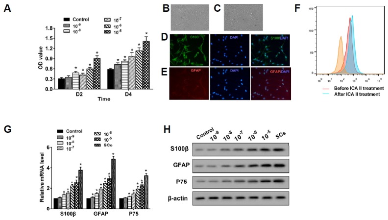
Fig. 1. ICA II promoted ADSCs’ proliferation and differentiation into SCs.

(A) ADSCs were treated with ICA II with a concentration of 10−9–10−5 mol/L and cell proliferation was determined through a CCK-8 assay at 48 h (D2) and 96 h (D4), respectively. *P < 0.05 vs. control. (B, C) After treatment with ICA II for 72 h, the morphology of ADSCs and the SCs differentiated from the ADSCs were obtained using a microscope (400×). (D, E) The expression of S100β and GFAP in the Schwann cells differentiated from the ADSCs was also confirmed by using the immunofluorescence staining method. (F) The percentage of S100β+GFAP+ cells was clearly elevated by ICA II in the process of differentiation from ADSCs to Schwann cells. (G) The mRNA levels of S100β, GFAP, and P75 in the ADSCs were quantified using qRTPCR. *P < 0.05 vs. control. (H) The expression of S100β, GFAP, and P75 proteins was analyzed through Western blot.
