
Figure 1. Adenoma colonoid appearance in culture.
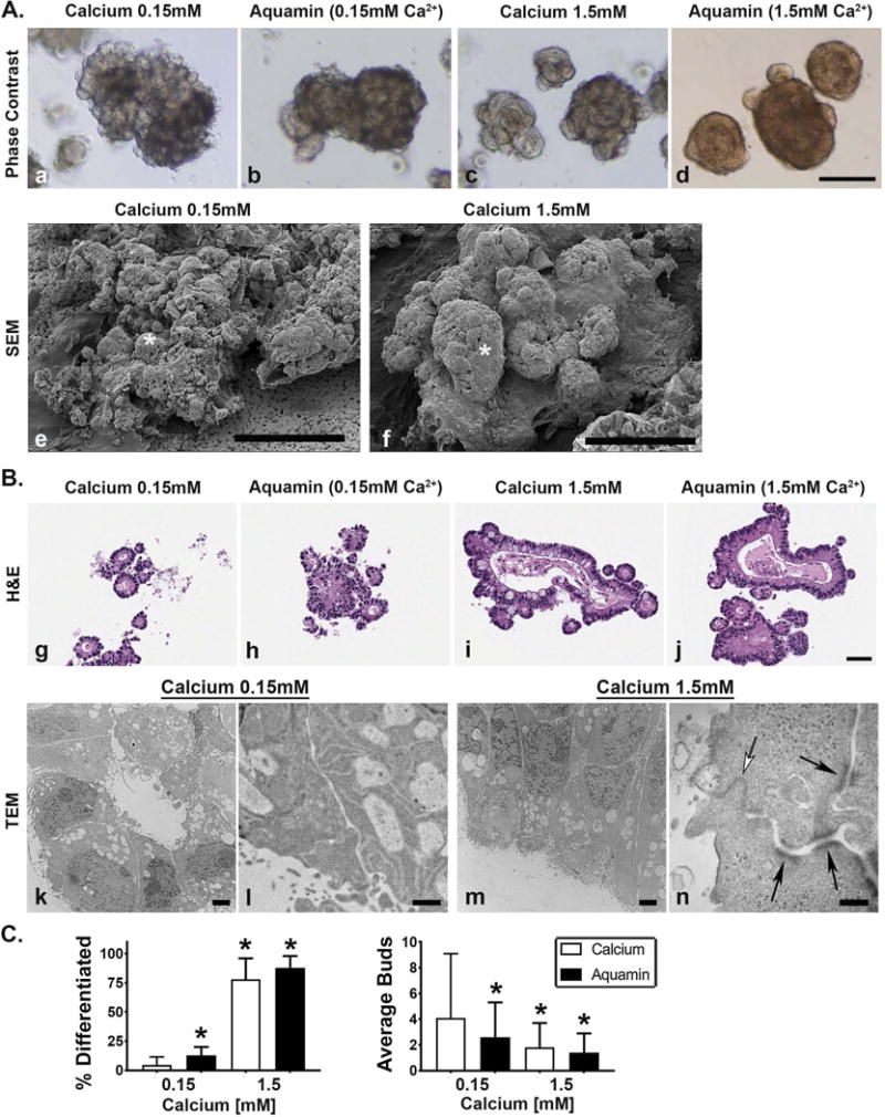
Figure 1A: Phase-contrast and scanning electron microscopy. At the end of the incubation period, virtually all of the adenoma colonoids maintained in 0.15 mM calcium consisted of a core structure with multiple tiny buds on the surface as indicated by phase-contrast microscopy (Figure 1, panel a). Colonoids maintained in 1.5 mM calcium (Figure 1, panel c) or treated with Aquamin® to provide either 0.15 mM (Figure 1, panel b) or 1.5 mM calcium (Figure 1, panel d) had a smooth surface with few buds. Bar=200μm. Scanning electron microscopic images confirmed the presence of multiple tiny buds growing out from the colonoid core structure under low-calcium conditions (asterisk) (Figure 1, panel e) and the presence of fewer but larger buds in the colonoid maintained in 1.5 mM calcium (asterisk) (Figure 1, panel f). Bars=100μm. Figure 1B: Histology and transmission electron microscopy. At the end of the incubation period, colonoids were examined under light-microscopy after sectioning and staining with hematoxylin and eosin. Under control conditions (Figure 1, panel g), tiny crypts of approximately 8-20 cells in cross section were seen. In the presence of 1.5 mM calcium alone (Figure 1, panel i) or with Aquamin® providing 1.5 mM calcium (Figure 1, panel j), larger crypts made up of columnar epithelial cells surrounding a large, often irregular-in-shape lumen were seen. Goblet cells were apparent. Colonoids treated with Aquamin® providing 0.15 mM calcium contained a mix of tiny crypts and larger structures (Figure 1, panel h). Bar= 50μm. When examined by transmission electron microscopy, the tiny crypts in low-calcium medium were found to consist of cuboidal cells surrounding a tiny central lumen. No desmosomes or tight junctures were seen (Figure 1, panels k and l). Under high-calcium conditions, the large crypts were made up of columnar epithelial cells around a larger lumen. Numerous desmosomes (black arrows) along the lateral surface connected by visible intermediate filaments and a tight-juncture at the apical surface (white arrow) were seen (m and n). Bar=2μm in Figure 1, panel k and m; Bar=500nm in Figure 1, panel l and =100nm in Figure 1, panel n. Figure 1C (Left panel): Percentage of individual colonoids with few tiny buds and smooth surface (phase-contrast). Means and standard deviations based on 20 fields per condition with 10-20 colonoids per field at 200X. Asterisks indicate statistical significance from control at p<0.05 level. Figure 1C (Right panel): Number of buds per core structure (hematoxylin and eosin sections). Means and standard deviations based on 5-10 high power fields per condition with 10-20 colonoids per field at 200X. Asterisks indicate statistical significance from control at p<0.05 level.