
Figure 4. STK11/LKB1 genomic alterations are enriched in LUAC with intermediate or high TMB that are negative for PD-L1 expression.
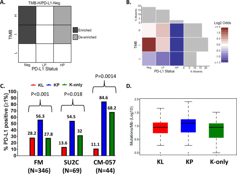
(A) PD-L1/TMB landscape matrix illustrating the enrichment analysis strategy in 924 LUAC samples with available CGP and PD-L1 expression (FM cohort). Enrichment of individual genomic alterations in PD-L1Neg; TMBI/H vs PD-L1HP; TMBI/H tumors was assessed using a one-sided Fisher’s exact test. (B) Heatmap of log-odds values reflecting the prevalence of STK11/LKB1 alterations in different cells of the PD-L1/TMB matrix. Alterations in STK11/LKB1 primarily cluster in TMBI;PD-L1Neg LUAC. (C) PD-L1 expression in the KL, KP and K-only subgroups in the FM (N=346), SU2C (N=69) and CM-057 (N=44) cohorts. A two-tailed Fisher’s exact test (computed from a 2×3 contingency table) was used to assess the significance of the association between group membership and PD-L1 expression status [PD-L1 positive (≥1%) or negative (0%)]. (D) TMB (Log10) in the KL, KP and K-only subgroups among 346 KRAS-mutant LUAC in the FM cohort.